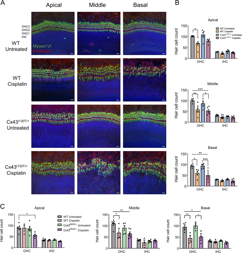
Fig. 3. Cisplatin causes hair cell loss in organotypic cochlear cultures from WT and Cx43I130T/+ mutant mice.
a Representative confocal images of organotypic cochlear cultures from WT and Cx43I130T/+ mutant littermate mice either untreated or treated with 20 μM cisplatin for 48 h. MyosinVI was immunolabeled to denote the hair cell body (green), phalloidin staining was used to demarcate actin (red), and Hoechst staining was used to locate the position of the nuclei (blue). b Quantitative analysis of hair cell counts in apical, middle, and basal regions of Cx43I130T/+ and WT littermate mouse cultures, either untreated or treated with 20 μM cisplatin. N values; WT untreated = 4, WT cisplatin = 5–6, Cx43G60S/+ untreated = 5, Cx43G60S/+ cisplatin = 4–5. c Quantitative analysis of hair cell counts in apical, middle, and basal regions of cultures for Cx43G60S/+ mutant mice and WT littermates. N values; WT untreated = 4–6, WT cisplatin = 3–4, Cx43G60S/+ untreated = 3–5, Cx43G60S/+ cisplatin = 3–6. OHC outer hair cell, IHC inner hair cell. Means represent mean ± standard error. Scale bars = 20 μm. Two-way ANOVAs were performed for each cochlear region with a subsequent Tukey’s post hoc test. *p < 0.05, **p < 0.01, ***p < 0.001.

