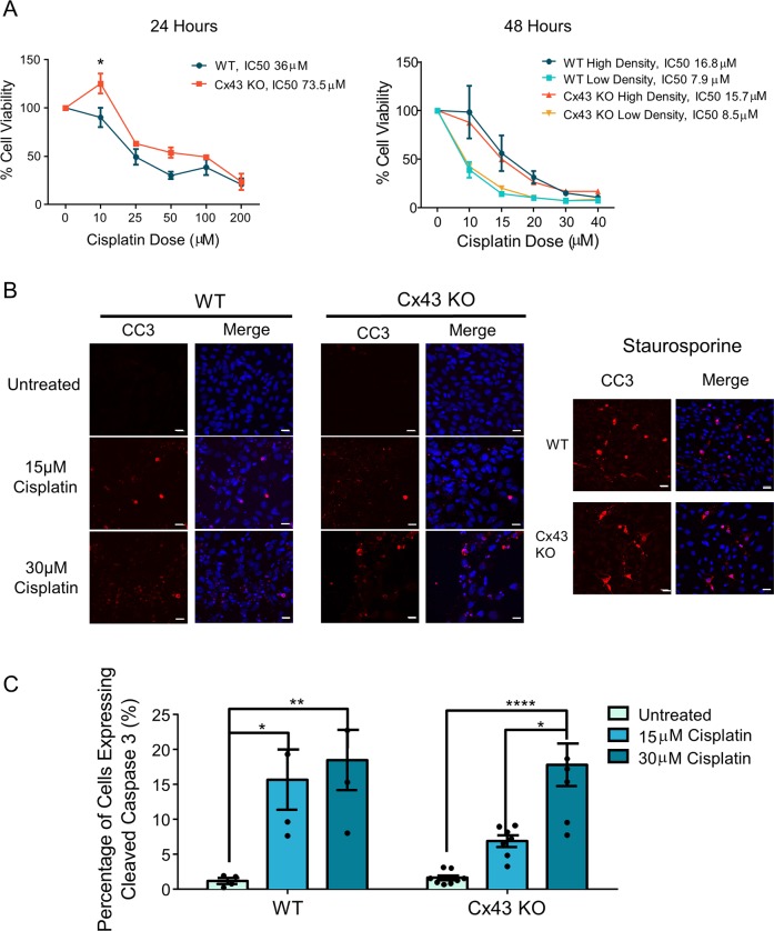
Fig. 5. WT and Cx43-ablated HEI-OC1 cells both have reduced cell viability after cisplatin treatment.
a Cell viability of HEI-OC1 cells at 24 and 48 h after cisplatin treatment. IC50s were calculated as the dose required to kill 50% of cells. b Representative confocal images of CC3 staining in control and cisplatin-treated cells. CC3 is denoted in red, and nuclei are in blue. Staurosporine-treated cells were used as a positive control. c Quantification of the percentage of CC3-positive cells showed a significant increase in the percentage of CC3-positive cells after cisplatin treatment. Bars represent mean ± standard error from four independent experiments comprised of two different Cx43 KO HEI-OC1 clones. N values; WT: N = 4, Cx43 KO: N = 7. Scale bars = 10 μm. Two-way ANOVA with Tukey’s post hoc test, *p < 0.05, **p < 0.01, ****p < 0.0001.

