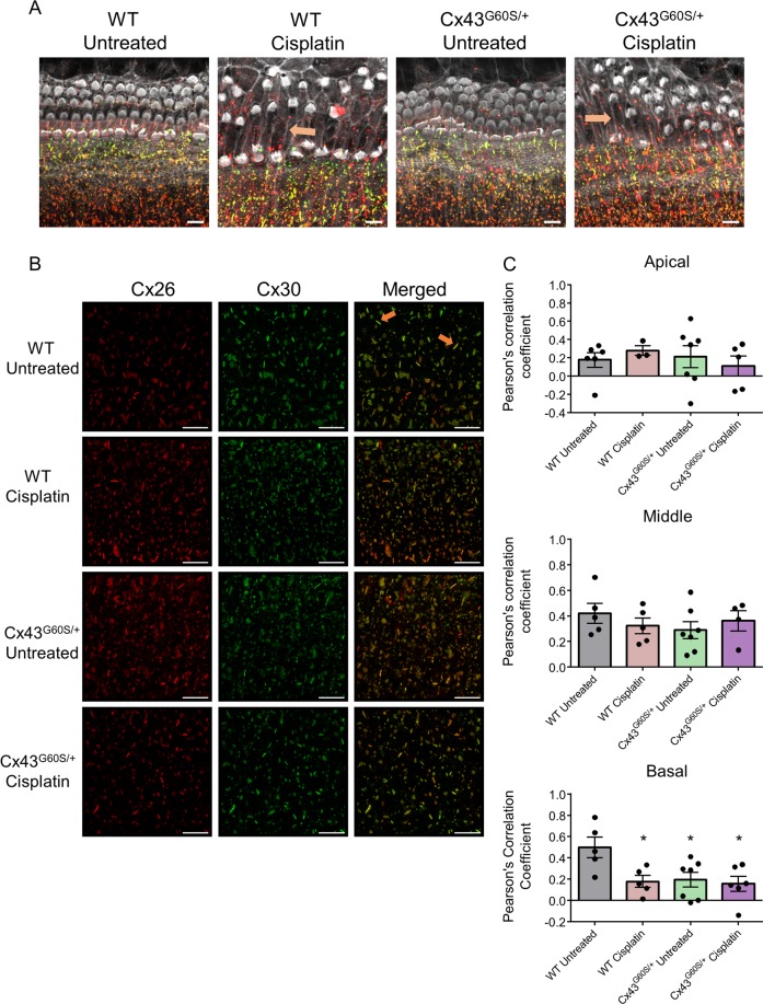
Fig. 6. Cisplatin treatment alters the spatial location of hair cells, causes supporting cell expansion, and induces the reorganization of Cx26 and Cx30 gap junctions.
a Representative confocal images of the basal region of organotypic cochlear cultures obtained from untreated and cisplatin-treated Cx43G60S/+ mutant mice and WT littermates. Orange arrows denote areas of pillar cell expansion. b High-magnification Airyscan images were acquired of the basal area from the inner sulcus region. Cx26 is denoted in red and Cx30 is in green. c Pearson’s correlation coefficient was quantified in apical, middle, and basal regions of organotypic cultures to measure Cx26 and Cx30 co-localization. N values; WT untreated = 5–6, WT cisplatin = 3–5, Cx43G60S/+ untreated = 6–7, Cx43G60S/+ cisplatin = 4–6. Means represent mean ± standard error. Scale bars = 10 μm. One-way ANOVA’s were performed for each cochlear region and subsequently a post hoc Tukey’s test was performed. *p < 0.05.

