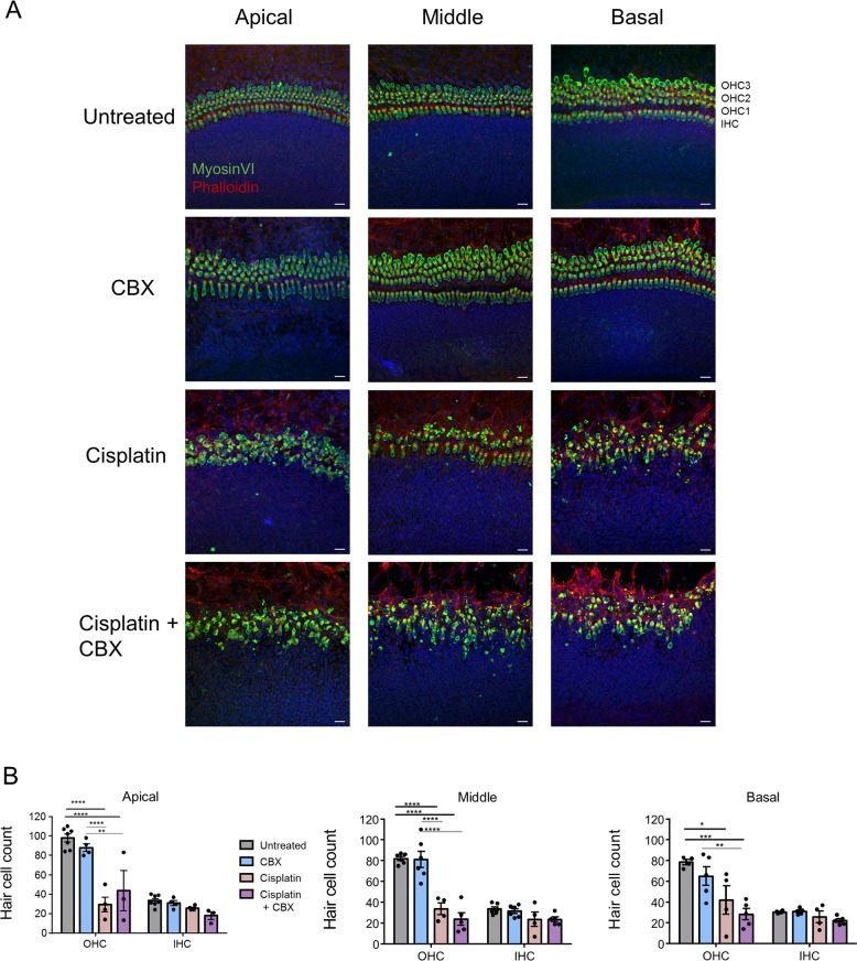
Fig. 8. Blocking gap junctions does not impact cisplatin-induced hair cell loss.
a Representative confocal images of hair cells in WT cultures that were untreated or treated with 100 μM carbenoxolone (CBX), 20 μM cisplatin, or 100 μM CBX and 20 μM cisplatin (cisplatin + CBX) for 48 h. MyosinVI is denoted in green, phalloidin is in red, and nuclei are in blue. b Quantification of hair cell counts from all four treatment groups at the apical, middle, and basal regions. N values; untreated = 4–7, CBX = 4–6, Cis = 4, Cis+ CBX = 3–5. OHC outer hair cell, IHC inner hair cell. Bars represent mean ± standard error. Scale bars = 20 μm. Two-way ANOVA’s and subsequent Tukey’s post hoc tests were performed for each region. *p < 0.05, **p < 0.01, ***p < 0.001, ****p < 0.0001.

