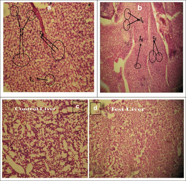
Fig. 8.
a Photomicrographs of histology of Control local tissue area stained with hematoxylin and eosin on day 15 after surgery captured by Leica Microscope at 40X respectively and f,c and I, represent spindle shaped fibroblast, collagen and Foreign body giant cells respectively b Photomicrographs of histology of Test local tissue area stained with hematoxylin and eosin on day 15 after surgery captured by Leica Microscope at 40X respectively and f,c and I, represent spindle shaped fibroblast, collagen and Foreign body giant cells respectively. c Photomicrographs of histology of liver of Test and control mice stained with hematoxylin and eosin on day 15 after surgery captured by Leica Microscope at 40X d: Photomicrographs of histology of kidney of control and test mice stained with hematoxylin and eosin on day 15 after surgery captured by Leica Microscope at 40X

