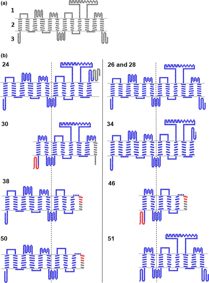
Figure 4.

(a) Predicted 2D structure of reference OATP1B1 (1: extracellular, 2: transmembrane, 3: intracellular) and (b) the predicted 2D structure of splice variants of OATP1B1, centered on the fourth intracellular loop (dashed line) of the reference structure for OATP1B1. The number of the splice variant is presented in the upper left corner of each structure. Red and blue: overlapping amino acid sequence with OATP1B1. Blue: overlapping structure OATP1B1.
