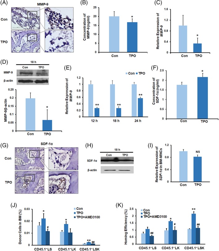
Figure 3.

TPO enhances HSPC homing by the downregulation of MMP‐9 expression and secretion. A, Immunostaining of femur sections with anti‐MMP‐9 antibody (bar = 100 μm). B, BM MMP‐9 levels in the recipient mice without BMT. C, Q‐PCR for MMP‐9 gene expression in the recipient BM cells. D, Western blotting of the recipient BM cells without transplantation using an antibody against MMP‐9. E, Q‐PCR for MMP‐9 gene expression of in vitro cultured BM cells. F, BM SDF‐1α levels in the recipient mice at 20 hours following a single dose of TPO or PBS injection. The BM samples were collected from the supernatants of BM cell suspensions that were flushed from two femurs using 1 mL PBS and were assayed with a SDF‐1α ELISA kit. G, Immunostaining of femur sections with an anti‐SDF‐1α antibody (bar = 100 μm). BM sections were prepared at 20 hours after TPO or PBS treatment. H, Western blotting of the recipient BM cells without transplantation using an antibody against SDF‐1α. I, Q‐PCR for SDF‐1α gene expression in the recipient BM cells. J,K, The donor cell percentages and homing efficiency analyses indicating that treatment with AMD3100 blocked the TPO‐enhanced homing of donor LSK cells. n = 3 each. *P < .05, **P < .01, compared with the control group; # P < .05, ## P < .01, compared with the TPO group. All recipient mice were lethally irradiated and received single‐dose TPO or PBS injections without BMT. BM, bone marrow; BMT, bone marrow transplantation; HSPC, hematopoietic stem and progenitor cell; MNC, mononuclear cell; PBS, phosphate‐buffered saline; Q‐PCR, quantitative polymerase chain reaction; TPO, thrombopoietin
