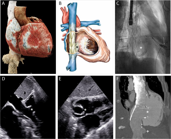
Figure 6.
Heterotropic transcatheter caval valve implantation. (A) 3D MSCT reconstruction of the vena cava inferior, the liver veins and the right heart cavities. (B) Schematic depiction of the NVT Tricento bicaval stenting device. (C) Fluoroscopic image of the implanted stent (projection: RAO 45). (D,E) Transthoracic echocardiographic imaging of the implanted device in his long and short axis from subxyphoidal at 30-day follow-up. (F) Depiction of the prosthesis and its relation to the right atrium and the hepatic vein in computed tomography. (Asterisk: valve element; arrow: leadless pacemaker; plus: hepatic vein).

