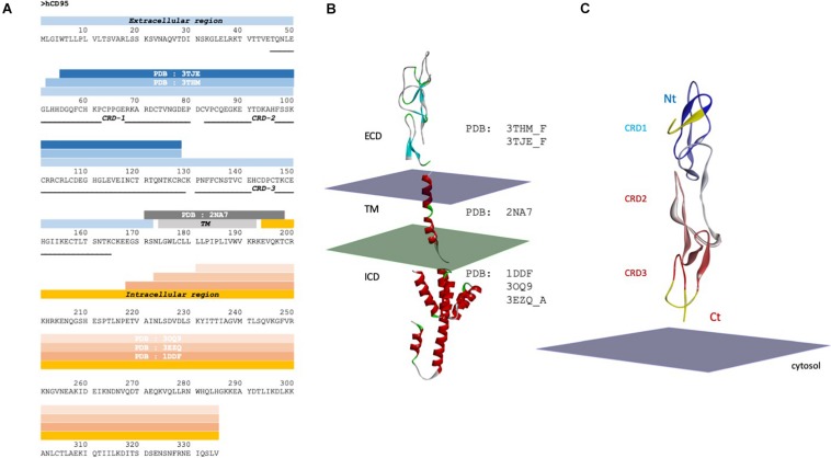
FIGURE 2.
CD95 sequence and structure. (A) Sequence of CD95 with solved 3D structures and corresponding PDB ID code. Blue, gray and orange strips represent the extracellular domain, the transmembrane domain and the intracellular region of CD95, respectively. CRD, cysteine rich domain; TM, transmembrane; ICD, intracellular domain; ECD, extracellular domain. (B) Domains of a monomeric CD95 whose structure has been experimentally solved. The plasma membrane is symbolized by two parallel planes, with the outer leaflet in purple and the cytosolic couleur in green. Note that the orientation toward membrane is a hypothesis. (C) Structure of the extracellular domain of CD95. Crystal structure of CD95 ECD domain (PDB:3TJE), colored according to the sequence order (blue to red, from Nt to Ct extremities). The yellow structure (amino acid residues N31 to D55) represents the gap in the crystal structure, which has been completed using CD40 homology. Nt: Amino-terminal region; Ct: COOH-terminal region.

