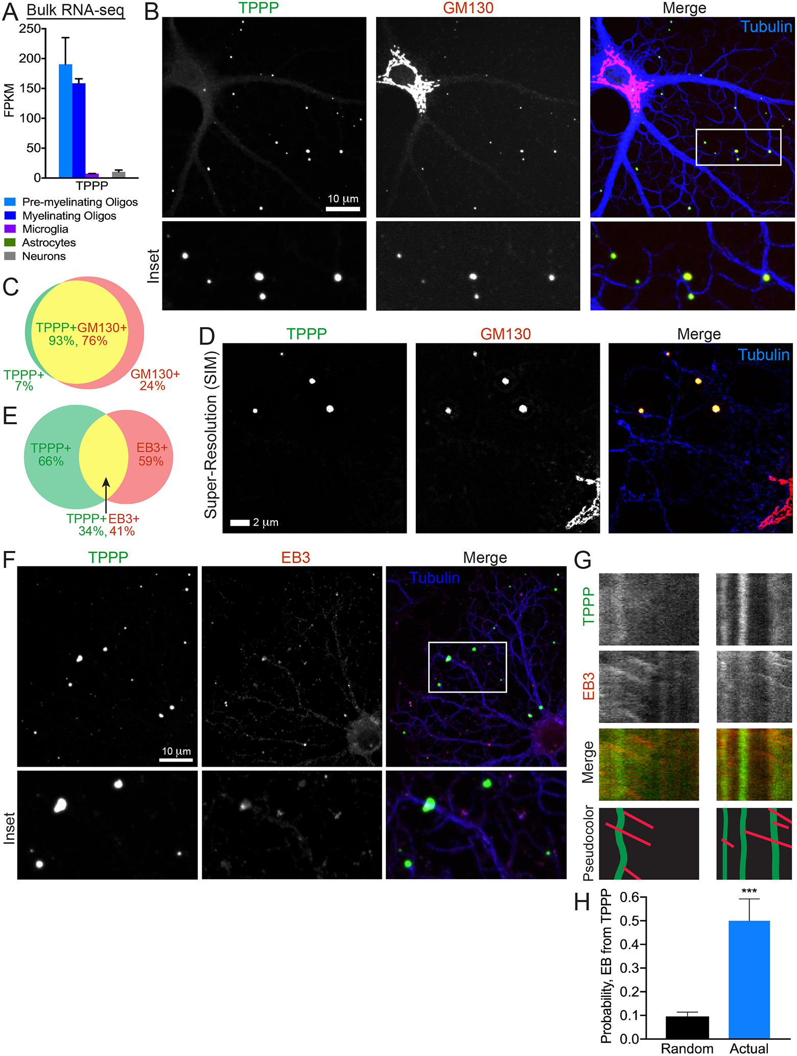
Figure 2. TPPP is a Specific Marker for Golgi Outposts.

(A) TPPP mRNA expression from RNA-seq of immunopanned mouse brain cells (Zhang et al., 2014). FPKM (frequency per kilobase million).
(B) Confocal micrograph of a rat DIV4 oligodendrocyte immunostained against TPPP, GM130, and tubulin.
(C) Venn diagram of colocalization relationship between TPPP and GM130 outside the cell body. 93% of TPPP+ puncta colocalize with GM130; 76% of GM130+ puncta colocalize with TPPP.
(D) Super-resolution (structured illumination) micrograph of a rat DIV4 oligodendrocyte immunostained against TPPP, GM130, and tubulin.
(E) Venn diagram of colocalization relationship between TPPP and EB1 outside the cell body. 35% of TPPP+ puncta colocalize with EB1; 41% of EB1+ puncta colocalize with TPPP.
(F) Confocal micrograph of rat DIV4 oligodendrocyte immunostained against tubulin, TPPP, and EB1.
(G) Kymographs from rat DIV1–2 oligodendrocytes expressing TPPP-GFP and mCherry-EB3.
(H) Comparison of actual rates of EB3-positive puncta arising from TPPP+ puncta versus probability based on chance. n = 9 cells, 3 biological replicates.
See also Figure S2.
