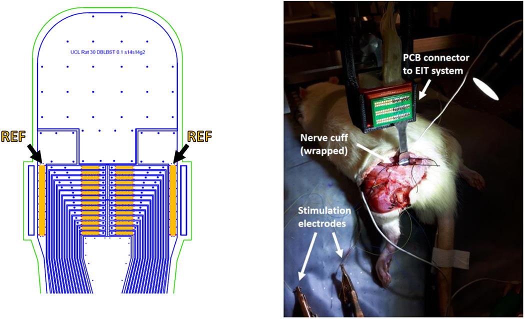
Figure 2.

Left: AutoCAD drawing of the top of the nerve cuff with electrodes (orange), outline of stainless steel tracks (blue) and outline of external cuff boundary (green). Arrows and labels indicate reference electrodes. Right: nerve cuff in an experimental setting, wrapped around the rat sciatic nerve.
