Figure 1.
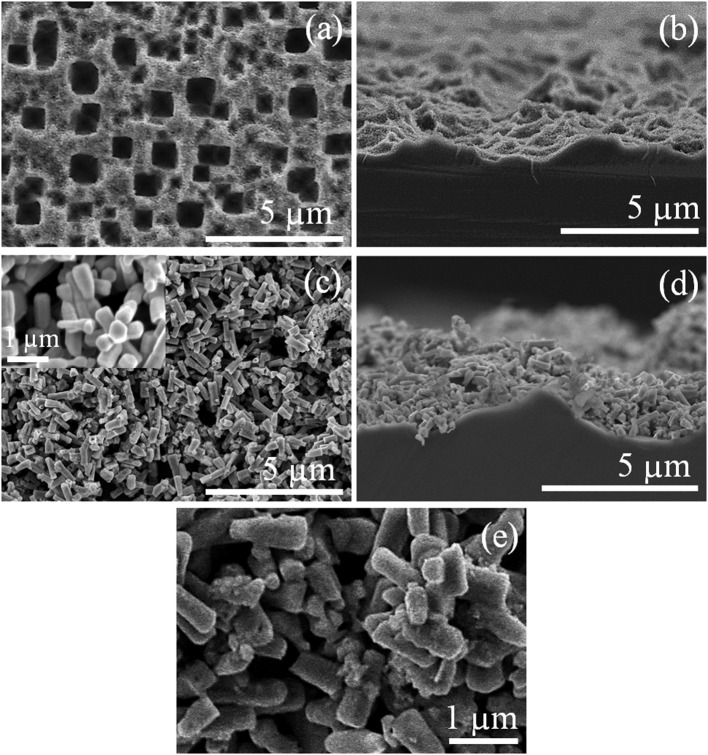
Scanning electron microscopy (SEM) micrographs of the pSi substrate (a) before and (c) after ZnO deposition (the inset shows an magnified area of the ZnO layer) and (b, d) their respective cross-section view; (e) ZnO–pSi hybrid structure after 1.0 μl nitrogen-doped carbon dots (NCDs) suspension deposition.

