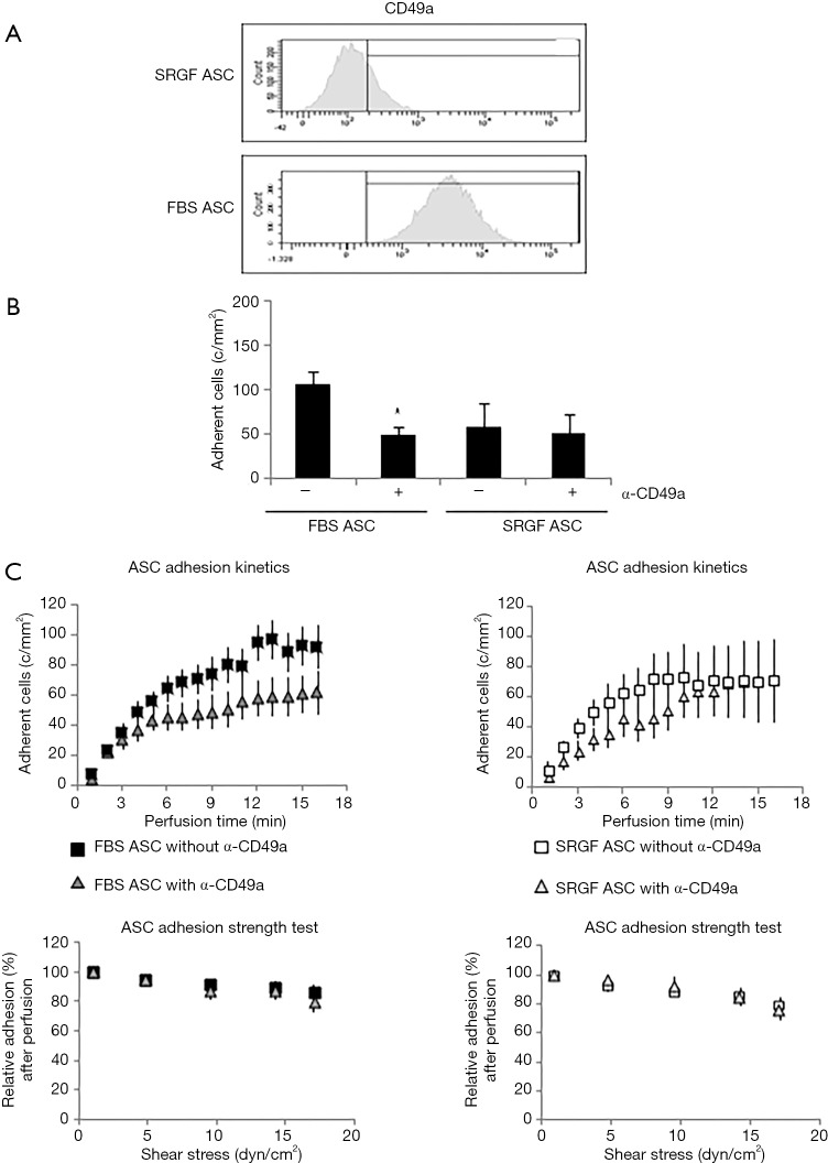
Figure 4.
Role of CD49a in FBS and SRGF ASC adhesion on collagen type I and fibronectin. (A) Representative example of CD49a expression analysis performed by flow cytometry in FBS and SRGF ASC. The threshold discriminating positively labeled cells was defined analyzing FBS or SRGF ASC in presence of the appropriate isotype control antibody (histogram not shown); (B) effect of anti-CD49a blocking antibody on FBS and SRGF ASC binding potential on collagen type I and fibronectin; (C) effect of anti-CD49a blocking antibody on FBS and SRGF ASC binding kinetics and adhesion stability on collagen type I and fibronectin. In ASC adhesion strength test reported in (C), density of adherent FBS or SRGF ASC immediately after perfusion in anti-CD49a added SM was expressed as 100% (first dot) and the fraction of adhering FBS or SRGF ASC along with the adhesion strength test was relatively expressed as percent. Assessments of CD49a expression (A) and function (B) were performed in FBS and SRGF ASC derived from all 5 patients. Perfusions were performed at 0.25 dyn/cm2 in standard medium (10% FBS α-MEM medium added with 2% bovine serum albumin); c/mm2, cells/mm2.

