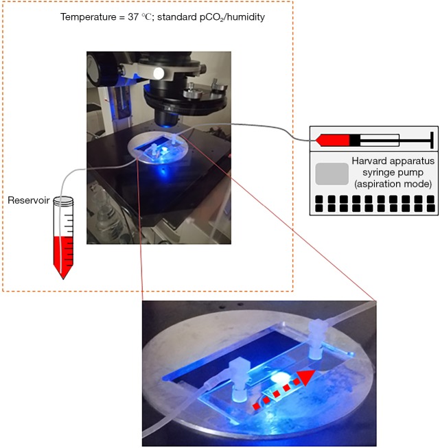
Figure S1.
Schematic representation of devices used to perform ASC adhesion and detachment assays. The parallel flow chamber slide (see photographic enlargement), was placed on an inverted epifluorescence microscope. Washed ASC after fluorescent labeling were resuspended with perfusion medium in a conical tube (Reservoir). In controlled environment (37 °C; standard atmospheric pCO2 and humidity) ASC were perfused in the parallel flow chamber through a small bore catheter. Flow was imposed by a syringe pump (Harvard apparatus). Adherent and detaching ASC were visualized by a digital camera and the number of adherent cells was estimated by image analysis approach. Timing of flow rate regulation and of digital image capture was reported in methods section and in Figure 1A. Dashed arrows represent the direction of flowing medium containing labeled ASC. Dashed orange box encloses devices placed under controlled environmental conditions.

