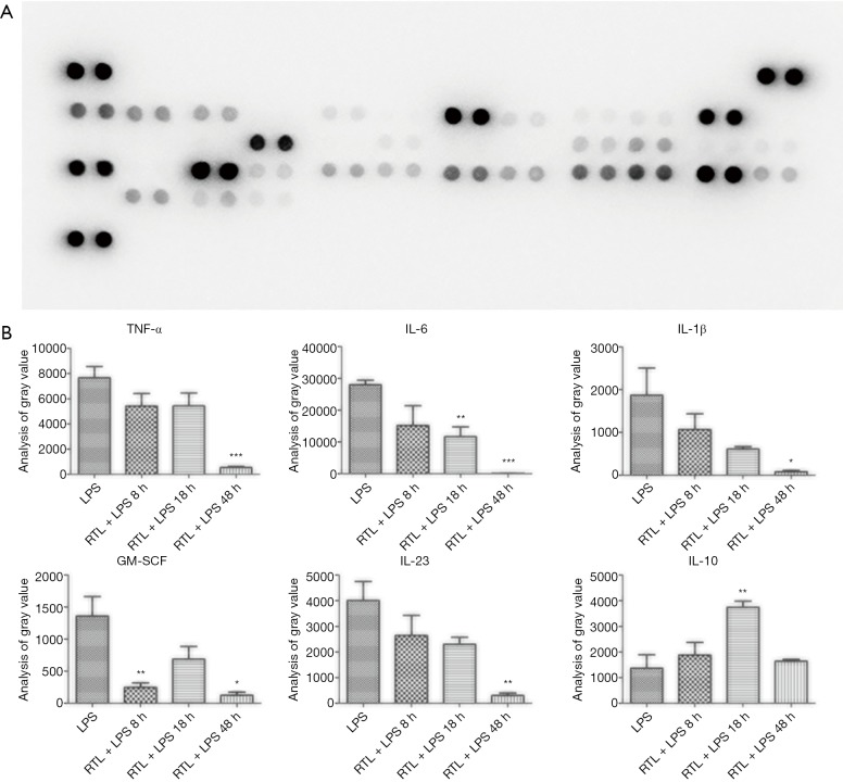
Figure 4.
The inhibition of ruxolitinib on cytokines in LPS-challenged mice. C57BL/6 mice were injected with ruxolitinib (0.67 mg/kg, i.p.) or vehicle (saline) 30 min before LPS injection (20 mg/kg, i.p.), and the serums were harvested in 8, 18, 48 h after LPS injection. (A) The membrane represented Cytokine Array Panel. Round black spots indicated production of different cytokines, and gray value of each cytokine was calculated. The results showed gray value of TNF-α9 (B), IL-6 (C), IL-1β (D), GM-SCF (E), IL-23 (F) with ruxolitinib treated decreased in 8, 18, 48 h respectively compared with LPS control (8 h). The gray value of IL-10 increased in 18 h after ruxolitinib intervented compared with LPS group. The values are the mean ± SD of 4 different samples. Statistical significance was assessed by the Student’s t-test and is represented as follows: *, P<0.5; **, P<0.01; ***, P<0.001 vs. LPS. RLT, ruxolitinib; LPS, lipopolysaccharide.

