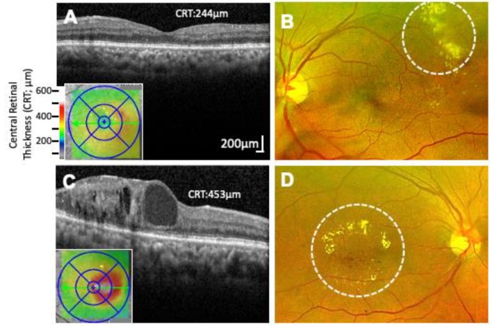
Figure 3.
Non-center and Center Involving Macular Edema. (A) Optical coherence tomography (OCT) image of the macular region of the retina showing non-center involving macular edema (Central retinal thickness, 244 µm). (B) Corresponding fundus photograph of (A) showing characteristic exudates formed outside of the macular region (marked by dashed circle). (C) OCT image of center involving macular edema with cystic changes within the retina (Central retinal thickness, 453 µm). (D) Corresponding fundus photograph of C showing characteristic hard exudates and edema formed inside the macula (marked by dashed circle).

