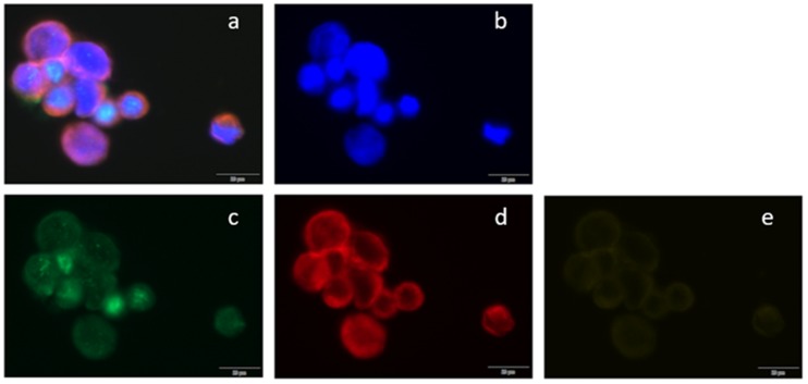
Figure 5.
Immunofluorescent staining of CTCs in metastatic breast cancer blood samples. Circulating tumor cells (CTCs) were scanned using (a) BX63 Upright Microscope; (a) a composite image of all channels, (b) 4’,6-diamidino-2-phenylindole (DAPI) counterstain (fluorescent blue), (c) estrogen receptor α stained with AlexaFluor488 (green), (d) cytokeratins 8, 18, and 19 stained with phycoerythrin (red), and (e) CD45 stained with AlexaFluor647 (yellow).

