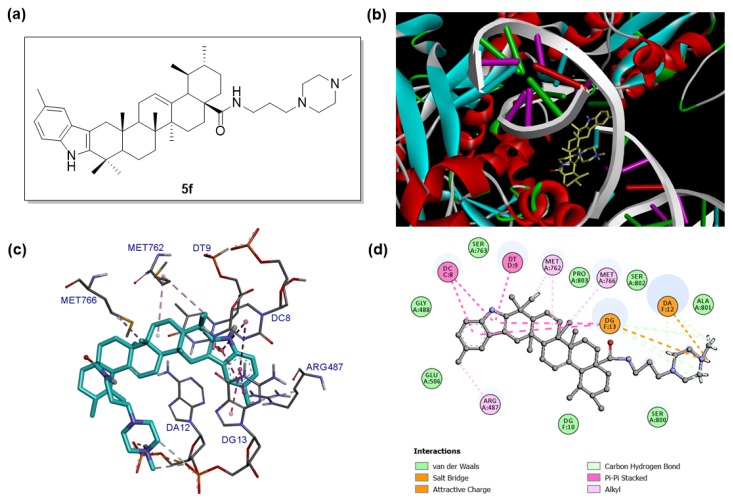
Figure 4.
Binding mode of compound 5f with human topoisomerase IIα in complex with DNA (PDB: 5GWK). (a) Molecular structure of compound 5f. (b) Binding pose of compound 5f within the active site of Topo IIα. Ligand is presented as stick models and colored by atom type, whereas the protein is presented as ribbons and DNA is presented as arrows (for backbone) and ladders (for base pairs). (c) Detailed docked view of compound 5f in the active site. The amino acid residues and base pairs are presented as sticks and colored by atom type, while the interactions are presented as dash lines. (d) Two-dimensional projection drawing of compound 5f docked into the active site of Topo IIα.

