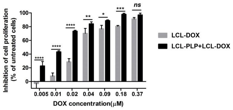
Figure 1.
Effects of LCL-PLP + LCL-DOX on B16.F10 cell proliferation. B16.F10 mouse melanoma cells co-cultured with murine macrophages were incubated with solutions of different concentrations of DOX (ranging from 0.005–0.37 µM) encapsulated in LCL (LCL-DOX) in the presence as well as in the absence of 410 μM PLP as LCL-PLP for 48 h. LCL-DOX, LCL-DOX-treated cells; LCL-PLP + LCL-DOX, cells incubated with solutions of different concentrations of LCL-DOX and 410 μM LCL-PLP. Data are shown as mean ± SD of three measurements and represented as percentages of proliferation inhibition compared with the proliferation of control cells. (ns, not significant (p > 0.05); * p < 0.05; ** p < 0.01; *** p < 0.001; **** p < 0.0001).

