FIGURE 2.
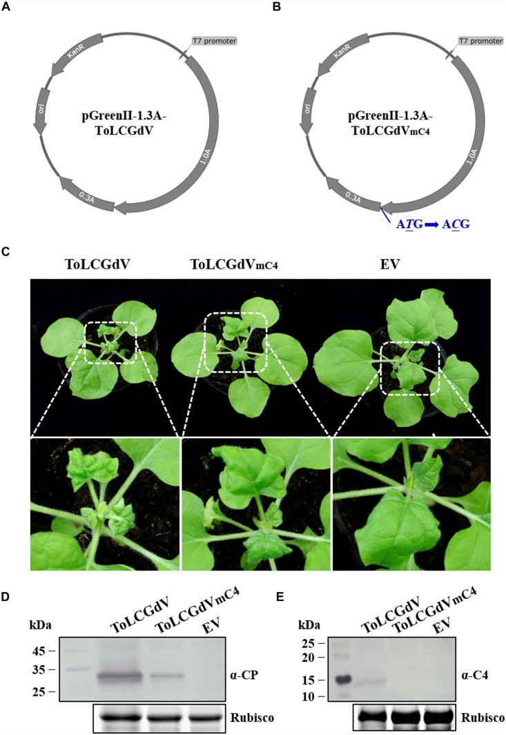
Construction of ToLCGdV and ToLCGdVmC4 infectious clones. (A) Schematic depiction of pGreenII-1.3A-ToLCGdV. Full-length and 0.3-time sequence of ToLCGdV were amplified and cloned into pGreenII vector (Hellens et al., 2000). KanR, kanamycin resistance. ori, replication initial origin. (B) Schematic depiction of pGreenII-1.3A-ToLCGdVmC4. Loss expression of C4 was made by replacing the start codon ATG with ACG, which has no effect on the expression of C1. KanR, kanamycin resistance. ori, replication initial origin. (C) Symptoms of N. benthamiana plants agroinfiltrated with pGreenII-1.3A-ToLCGdV or pGreenII-1.3A-ToLCGdVmC4. Agrobacterium strains harboring pGreenII-1.3A-ToLCGdV or pGreenII-1.3A-ToLCGdVmC4 were infiltrated into the leaves of 5–6 leaf-stage N. benthamiana. Photos were taken at 13 dpi. The bottom panel shows the magnification of the white dotted frame. EV, empty vector. (D) Western blot detection of virus accumulation in the infected plants. The upper leaves were taken at 13 dpi and subjected to western blot detection with anti-ToLCGdV AV1 (CP) antibody. Rubisco shows equal sample loading. Numbers on the left indicate molecular weight. (E) Western blot detection of C4 in the upper infected leaves. Leaves were taken at 13 dpi to perform western blot with anti-C4 antibody. Rubisco is used as equal loading. Numbers on the left indicate molecular weight.

