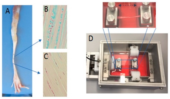
Figure 8.
Exemplary images of the tendon and the bioreactor used in the study. (A) Tendons of the flexor digitorum longus were extracted from rat legs with two different areas of traction and gliding and stained with an Alcian blue to indicate the varying expression of sulfated glycosaminoglycans (sGAG) in different areas of the tendon (gliding (B) and traction (C)). (D) The image shows the in vitro bioreactor that was used, in which the tendon was fixed between two pads.

