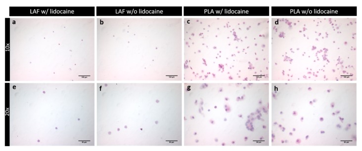
Figure 2.
This figure presents the lysed SVF of the lipoaspirate of the fluid (LAF) and fatty portion (PLA), which was later used for flow cytometry. Slides were observed in a light microscope. In (a,e), the LAF w/lidocaine, and in (b,f), the LAF w/o lidocaine is seen in 10× and 20× magnification. In panel (c,g), PLA w/lidocaine, and in (d,h), PLA w/o lidocaine is shown in 10× and 20× magnification.

