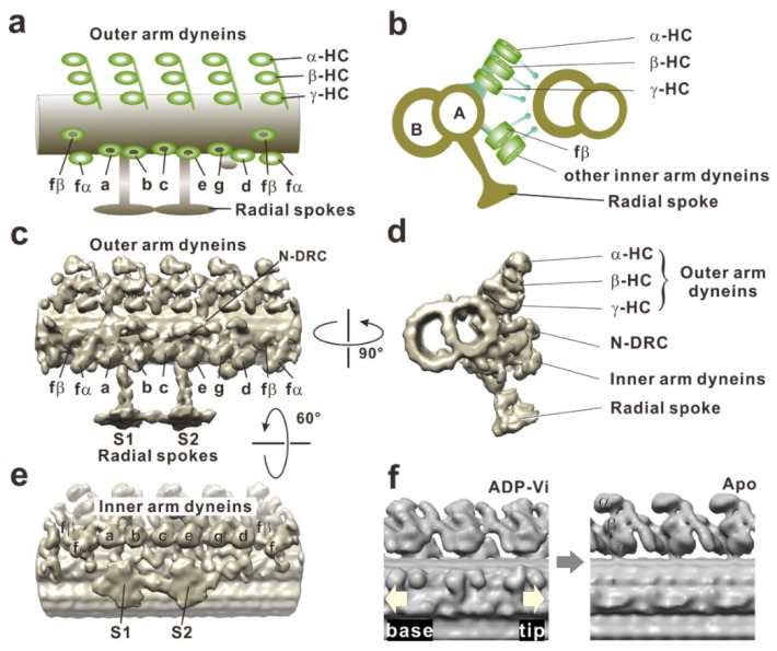
Figure 4.
Cryo-electron tomographic images of a doublet microtubule in a Chlamydomonas flagellum. (a) A schematic drawing of the dynein arms and doublet microtubule in the longitudinal section. Three heavy chains of outer arm dyneins (α, β, and γ) stack on top of each other, and four such trios align with the 96-nm periodicity. The left and right sides indicate proximal (base) and distal (tip) sides, respectively. (b) A schematic drawing of the doublet microtubules in the cross-section viewed from the flagellar base to the tip. (c) Surface-rendered representations of a doublet microtubule in a longitudinal section on the basis of cryo-electron tomography (EMD-2116). Dynein arms arrayed on the doublet microtubule are viewed from the adjacent doublet microtubule. A structure connects two adjacent doublet microtubules is the nexin-dynein regulatory complex (N-DRC). There are two radial spokes (S1 and S2). (d) Cross-section viewed from the proximal (from the cell body) to the distal side. The image clearly shows the three rows: outer dynein arm, inner dynein arm, and N-DRC. (e) Longitudinal section of the doublet microtubule viewed from the central pair apparatus. The arrangement of the inner arm dyneins is clearly visible. Eight heavy chains of inner arm dyneins (a, b, c, d, e, fα, fβ, and g) appear to form four pairs: fα–fβ, a–b, c–e, and g–d. (f) Conformational changes of outer arm dyneins coupled with nucleotide states were observed in the comparison of two tomography images, EMD-1696 and EMD-1697, from Movassagh et al. (2010) [47].

