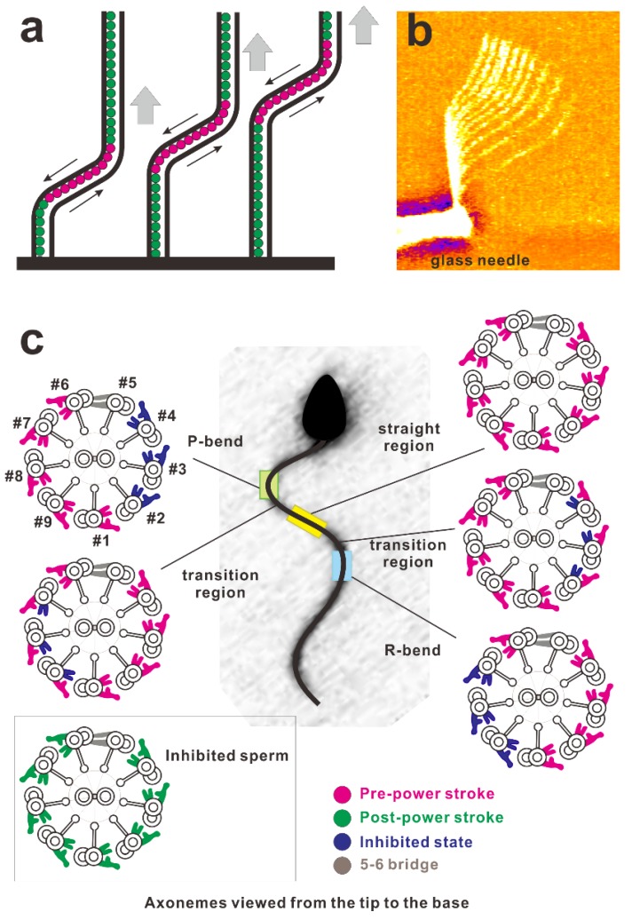
Figure 5.
(a) Schematic of the propagation of a bend in the axoneme. Red circles indicate the active dynein arms generating shear between the microtubules and green ones indicate inactive dynein arms. A bend activates dynein arms and propagates toward the tip. (b) Overlaid video images of a demembranated flagellum spontaneously propagating the bend applied near the base using the rapid movement of the glass needle (courtesy of Dr. Shingyoji). Soaking a demembranated flagellum in low concentrations of Mg-ATP (e.g., 2.5 μM) ceases its beating. Rapid movement of the microneedle generates a bend of the flagellum at the region near the base. This bend propagates toward the tip with a constant bend angle [32]. (c) Asymmetric distributions of dynein arms in the active state coupled with the bend revealed by electron tomography. (This diagram was redrawn with modifications according to Lin and Nicastro (2018) [48].) The sea urchin sperm flagellum during its swimming was rapidly frozen and observed using cryo-electron tomography. In this figure, the cross-section of the axoneme was drawn as the view from the distal (flagellar tip) to proximal (cell body) ends of the flagellum. The configuration of dynein molecules was categorized into four states and the distribution was examined using the tomography results. For further details of the observations, consult Lin et al. (2018) [48], Hastie et al. (1991) [100], or King (2018) [101]. Asymmetric distributions of the dynein states are shown to be coupled with bends.

