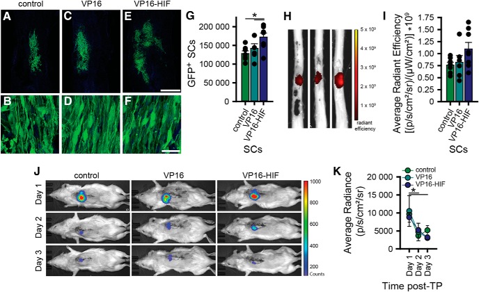
Figure 4.
HIF increases the survival of transplanted cells. This is detectable histologically by stereological quantification, but not by ex vivo fluorescent imaging of spinal cords or in vivo bioluminescent imaging; 2 × 10⁶ GFP-luc SCs were transplanted into the injured spinal cord 7 d post-SCI [control (n = 7), VP16 (n = 6), or VP16-HIF (n = 8)]. Seven days post-TP, transplant survival was quantified by histology (A–G) and ex vivo fluorescent imaging of spinal cords for GFP (H, I). In a subset of the rats [control (n = 3), VP16 (n = 4), or VP16-HIF (n = 4)], transplant survival was assessed by performing daily in vivo bioluminescent imaging on days 1–3 post-TP (J, K). Representative images of the transplants and the cells within the transplants 7 d post-TP are shown for each group (A–F). More SCs survive when they express VP16-HIF, as determined by stereology (G). Results of quantification of the cell number by stereology (G). Images of representative spinal cord showing radiant efficiency of the ex vivo imaging for transplant GFP fluorescence 7 d post-TP (H). Quantification of radiant efficiency (I). Images of photon counts for bioluminescence activity of the transplanted cells for the first 3 d post-TP for each cell type transplanted (J) and quantification of light emitted (average radiance; K). Mean ± SEM; *, G, control versus VP16-HIF, p = 0.0025; VP16 versus VP16-HIF, p = 0.027; K, d1 versus d2, p = 0.01; d1 versus d3, p = 0.031. Figure Contributions: Caitlin Hill, Brian David, Jessica Curtin, and David Goldberg performed the experiment. Caitlin Hill and Brian David analyzed the data.

