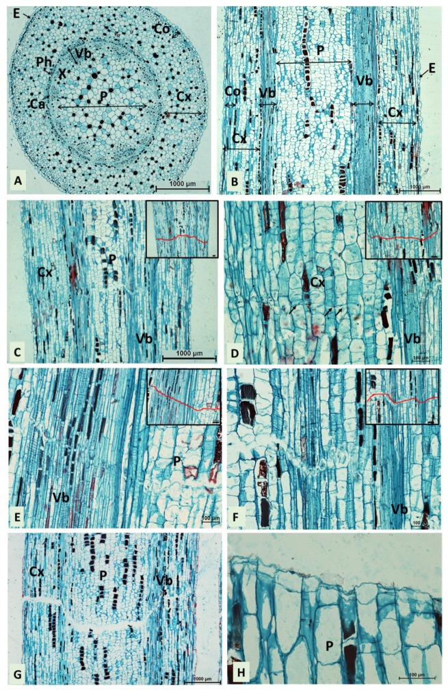
Figure 2.
Microscopy investigation of the secondary abscission zone in the middle of stem internodes induced by IAA 0.1% (longitudinal sections). (A,B) Anatomical details of the stem in the middle of internodes not treated with hormones (control) stained with safranin-fast green. (A): a cross-section; (B): a longitudinal section; CA–cambium; Cx–cortex; E–epidermis; CO–collenchyma; P–pith; PH–phloem; VB–vascular tissues; X–xylem. (C) the beginning of the formation of an abscission zone, cell separation visible in pith, photographed on the eighth day after treatments; (D) the formation of an abscission zone in the cortex; (E) cell separation in the cortex and pith; (F) the formation of an abscission zone in the vascular tissue region; (G) cell separation visible in all tissues of the stem internode; (H) the secondary abscission completed, photographed on the ninth day after treatments; (C–F) the insert displays the outline of the creation of abscission zones (red); bars in the inserts represent 100 µm; Cx–cortex; N–nucleus; P–pith; VB–vascular tissues. Arrows denote cell-plate forming between daughter cells.

