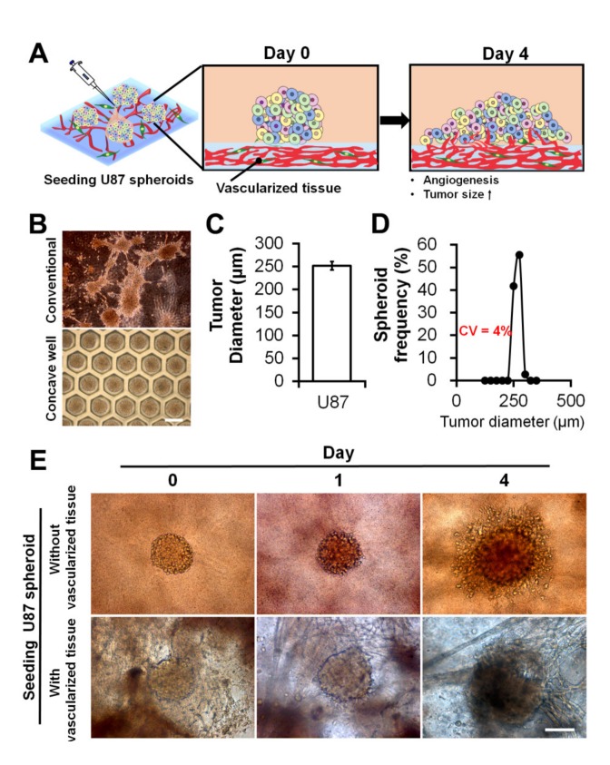
Figure 4.
Uniform-sized U87 MCTSs and their seeding onto GAF hydrogel layers with and without preformed vascularized tissues. (A) Schematic describing the morphological changes and angiogenesis of MCTSs seeded onto the vascularized tissue. (B) Optical images of MCTSs formed on petri dishes and concave wells on day three. The scale bar represents 400 μm. (C) Diameter and (D) size distribution of MCTSs formed in concave wells. (E) Optical images of MCTSs seeded onto GAF hydrogel layers with and without preformed vascularized tissues on days zero, one, and four. The layer with vascularized tissues was constructed by incubating the bioprinted GAF hydrogel encapsulating HUVECs and LFs for seven days in the media, and the layer without vascularized tissues was constructed by printing the bare GAF hydrogel. The scale bar represents 200 μm.

