Figure 2.
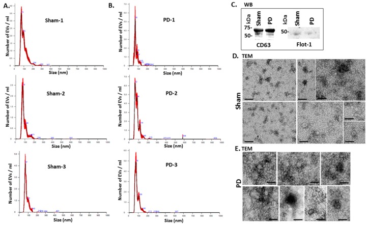
EV characterisation from rat plasma. (A) Representative Nanosight graphs showing nanoparticle tracking analysis (NTA) analysis of plasma EV profiles from sham-treated rats (Sham; n = 3). (B) Representative Nanosight graphs showing NTA analysis of plasma EV profiles from the pre-motor PD rat models (PD; n = 3). (C) Western blotting (WB) confirms that rat plasma-EVs are positive for the EV-specific markers CD63 and flotillin-1 (Flot-1); the molecular weight standard is indicated in kilo Daltons (kDa). (D,E) Transmission electron microscopy (TEM) images showing EVs isolated from sham (D) and pre-motor PD model (PD; (E)) rat plasma, revealing typical EV morphology; composite images are shown and the scale bar represents 50 nm in all images. In the NTA curves the black line represents the mean of the 5 repetitive readings per individual sample and the red line represents standard error (+/−) between those same 5 readings per sample. Each treatment group was measured in 3 biological replicates (sham; PD = pre-motor PD models).

