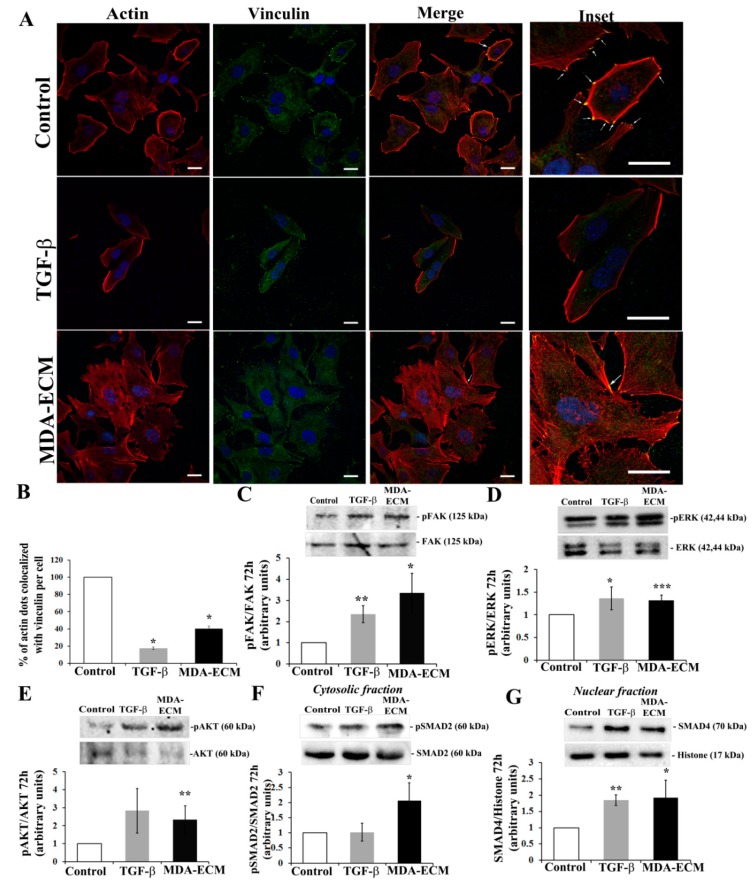
Figure 6.
Interaction with MDA-ECM triggered integrin and TGF-β-associated signaling pathways in MCF-7 cells. (A) MCF-7 and MDA-MB-231 cells were cultured in standard conditions on coverslips for 72 h, and decellularized ECMs were obtained, as described in Methods. For double staining of F-actin and vinculin (indicated by the white asterisks), cells were marked with rhodamine-conjugated phalloidin (red) and anti-vinculin antibody (green), and nuclei were stained with DAPI. Representative images were captured at 60× magnification. Focal adhesion assembly in control is highly visible in the section at high magnification (insets-right column). Scale bar: 20 µm. (B) Representative graph showing the percentage of colocalization of vinculin and actin in comparison to the control MCF-ECM. Cell lysates were immunoblotted with (C) anti-pFAKTyr397 and anti-FAK, (D) anti-pERK1/2 and anti-ERK1/2, (E) anti-pAKTSer473 and anti-AKT, (F) anti-pSMAD2 and anti-SMAD2, and (G) anti-SMAD 4 and anti-histone (H3) antibodies. The results are shown as the fold increase compared to the MCF-ECM group, calculated from 3 individual experiments (* p < 0.05; ** p < 0.01; *** p < 0.001).

