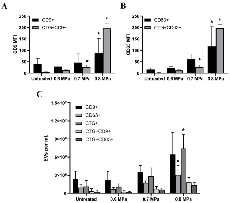
Figure 3.
Extracellular vesicles (EVs)-containing supernatants of HUVECs after CTG and USMB treatments measured using flow cytometry. EVs were captured by anti-CD9 and anti-CD63 beads in the supernatants of CTG-stained HUVEC followed by USMB treatment. These EVs were stained with anti-CD9 and anti-CD63 detection antibodies. The MFI of CD9 and CD63 levels in untreated and USMB treated samples (0.6, 0.7 and 0.8 MPa) were quantified using flow cytometry (A,B). These captured CD9 EVs and CD63 EVs carried CTG (A,B). Samples were measured in triplicate and these were data from two independent experiments. The same samples were measured using a micro flow cytometer. Anti-CD9 and -CD63 were used to detect EVs bearing CD9 and CD63 antigens present in the cell supernatants (C). Total EVs carrying CTG and EVs exposing CD9 or CD63 which carried CTG were also quantified in the same samples (C). Results are reported in EVs per mL sample. These were data from two independent experiments. As controls, EVs without CTG were used and all results presented have been corrected from these controls (A–C). Statistical analysis was performed using one-way ANOVA followed by Tukey’s test. The p value < 0.05 was considered significant (*).

