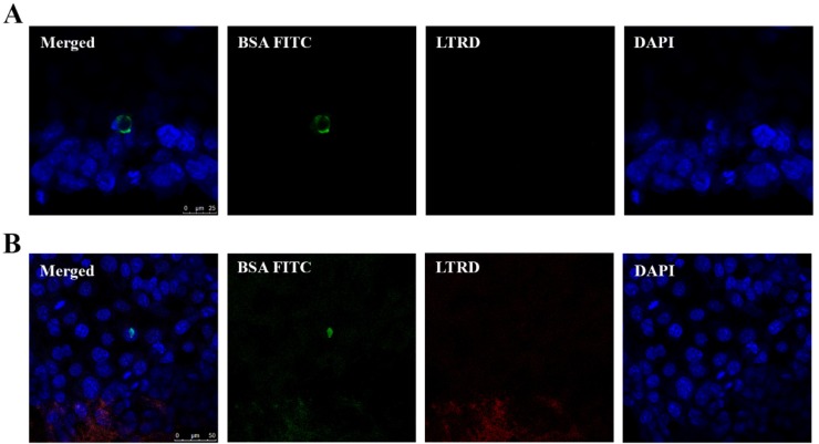
Figure 7.
Uptake of EVs containing BSA FITC by FaDu cells. FaDu cells were co-cultured with EVs containing BSA FITC (undiluted). After 4 h, cells were washed with acid wash buffer followed by PBS and fixed prior to staining. LTRD probe (red) was used to stain lysosome and DAPI (blue) to stain the nucleus. Merged images show the uptake of BSA FITC by FaDu cells. BSA FITC signal (green) seems to be present in the cell cytoplasm (A, merged) or in the vicinity of the cell nucleus (B, merged).

