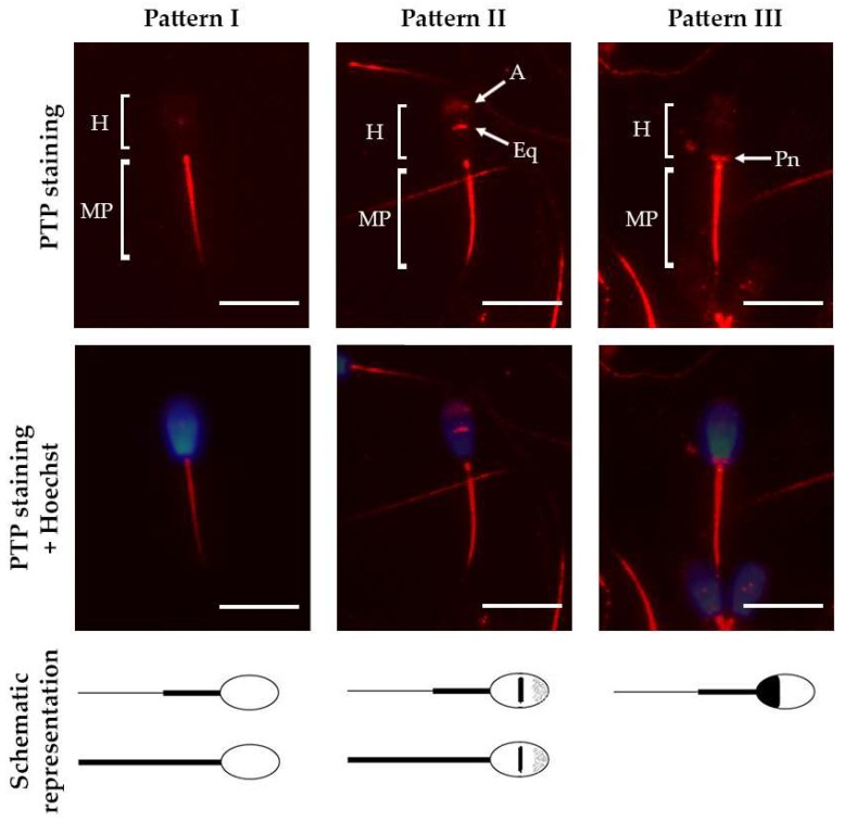
Figure 1.
Representative micrographs of the three PTP patterns in bull spermatozoa that were detected employing immunolabeling (red). Nuclei were counterstained with Hoechst 33342 (blue). Below the micrographs, a schematic representation of each pattern is shown. Pattern I: staining at the midpiece (MP) and/or the whole flagella; pattern II: staining at the acrosomal region (A), the equatorial region (Eq), the midpiece and/or the whole flagella; pattern III: staining at the post-nuclear region (Pn) and the midpiece. Scale bar represents 10 µm.

