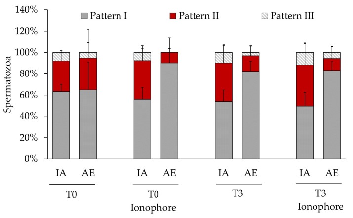
Figure 3.
Distribution of protein tyrosine phosphorylation in bull spermatozoa showing intact acrosome (IA) or acrosomal exocytosis (AE). Spermatozoa were analyzed after a treatment with or without ionophore A23187 at time 0 (T0) or after 3 h (T3) of incubation under capacitating conditions. Values of each pattern are represented as means (%) ± SD, n = 6.

