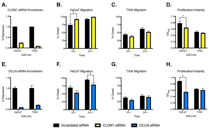
Figure 5.
Migration and proliferation/viability of skin and oral keratinocytes after CLDN1 or OCLN knockdown. For migration assays, HaCaT and TIGK cells were transfected with claudin 1 or occludin small interfering RNA (siRNA). The transfected cells were cultured in 2-well silicone culture inserts with a defined cell-free gap as described in the Materials and Methods section. Cell migration was documented by a digital camera 18 and 24 h later. For proliferation/viability assays, 5 × 10³ cells/well of claudin 1 or occludin siRNA-transfected HaCaT and TIGK cells were plated in a 96-well plate. A 3-(4,5-dimethylthiazol-2-yl)-5-(3-carboxymethoxyphenyl)-2-(4-sulfophenyl)-2H-tetrazolium, inner salt (MTS) proliferation assay was performed to record OD490 values corresponding to the numbers of live cells using a spectrophotometer. (A) CLDN1 knockdown in HaCaT and TIGK cell lines. (B,C) Migration after CLDN1 knockdown in HaCaT and TIGK cells, respectively. (D) Proliferation/viability after CLDN1 knockdown. (E) OCLN knockdown in HaCaT and TIGK cell lines. (F,G) Migration after OCLN knockdown in HaCaT and TIGK cells, respectively. (H) Proliferation/viability after OCLN knockdown. Black = scrambled siRNA, yellow = CLDN1 siRNA, blue = OCLN siRNA; n = 3 replicates, * p < 0.05.

