Figure 5.
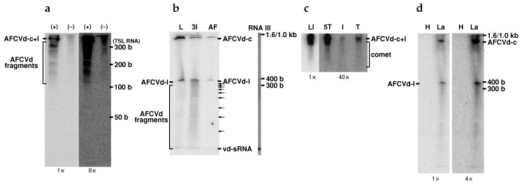
Figure 5. Northern blot analyses of shorter-than-unit-length degradation products of AFCVd in developing and germinating tobacco pollen: (a) 10 μg of 2 M-LiCl-soluble RNA isolated from AFCVd-infected pollen at developmental stage 3 was analyzed in 12% acrylamide gel containing 3 M urea and hybridized to strand-specific AFCVd RNA probes for detection of either (+) or (−) AFCVd sequences, as designated on top of the gel. (b) Analysis of 10 μg of 2 M-LiCl-soluble RNA isolated from AFCVd-infected tobacco leaves (L) and pollen from stage 3 (3I) using discontinuous-pH acrylamide gel electrophoresis. Samples were applied in 8% denaturing gel containing 8 M urea, hybridized with (−) AFCVd RNA probe for detection of (+) chains, monomeric circular (AFCVd-c) and linear (AFCVd-l) viroid. The arrows on the right indicate stronger degradation fragments of AFCVd from pollen stage 3. AF, partly purified AFCVd isolated using PEG fractionation of LiCl-soluble RNA. (c) Analysis of “comets” from tobacco infected leaves and pollen infected with AFCVd on 1.5% agarose gel; viroid-specific RNA was detected using hybridization to [32P]dCTP-labeled AFCVd cDNA. LI, 10 μg total RNA from infected leaves; 5T, I and T, 30 μg LiCl-soluble RNA from 35S:AFCVd vector-transformed pollen stage 5, infected germinating pollen and 35S:AFCVd vector-transformed germinating pollen, respectively. (d) Analysis of LiCl-soluble RNA from healthy (H) and vector pLAT52:AFCVd-transformed germinating pollen under similar gel conditions as in (b). After electrophoresis, nucleic acids in gels (a,b,d) were electroblotted to positively charged nylon membranes; a capillary blot was performed on Biodine A nylon membrane for gel (c). Positions of RNA markers are indicated on the right sides of the gels. Gel strip with RNA III marker was silver stained. As marker of 300 base RNA, position of 7SL RNA is indicated in (a). Positions of “comets” zones and cones where viroid RNA fragmentation is seen are indicated in this figure. Relative screen intensifications are given on the bottom of gels.

