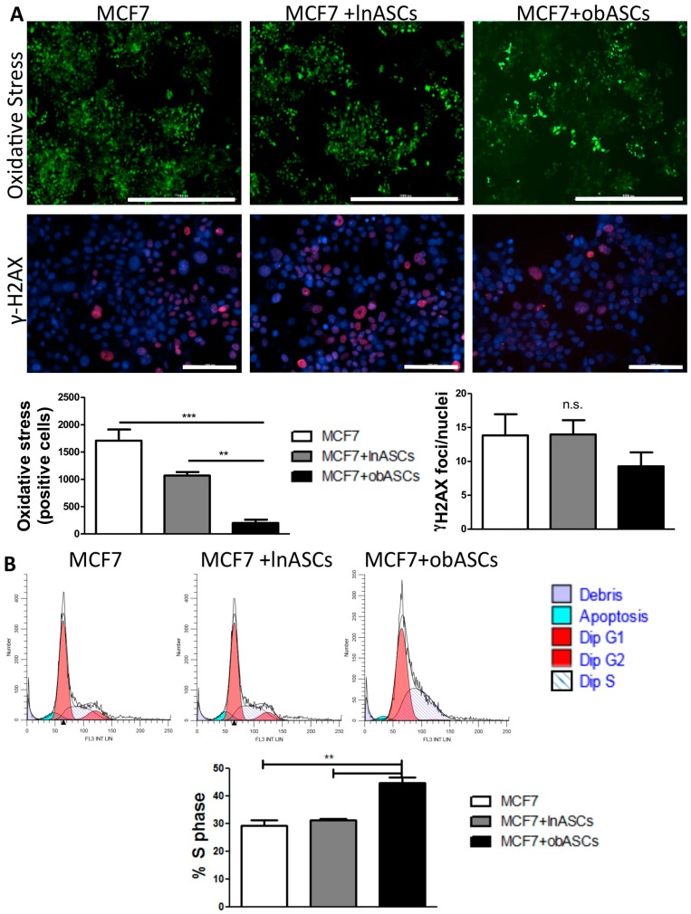
Figure 2.
obASCs decrease oxidative stress in breast cancer cells after radiation but have no effect on DNA damage. (A) DCFDA, which becomes fluorescent by reacting with oxidative species, was used to quantify number of cells undergoing oxidative stress after radiation. Quantification demonstrates that there is a significant decreased in cells that were co-cultured with obASCs undergoing oxidative stress. Evaluation of double stranded DNA breaks via immunofluorescent γ-H2AX staining with Hoechst 33342 nuclear counterstain revealed no difference in DNA damage after radiation between groups. Scale bar represents 1000 μm in the upper panel and 100 μm in the lower panel. (B) Cell cycle analysis using propidium iodide (PI) staining and ModFit software demonstrates percent of cells in the different phases of the cell cycle. Quantification reveals increased percent of diploid cells in S-phase after transwell co-culture with obASCs. Values reported are the mean of three independent experiments each performed in triplicate. Bars, ± SEM. ** p < 0.01, *** p < 0.001.

