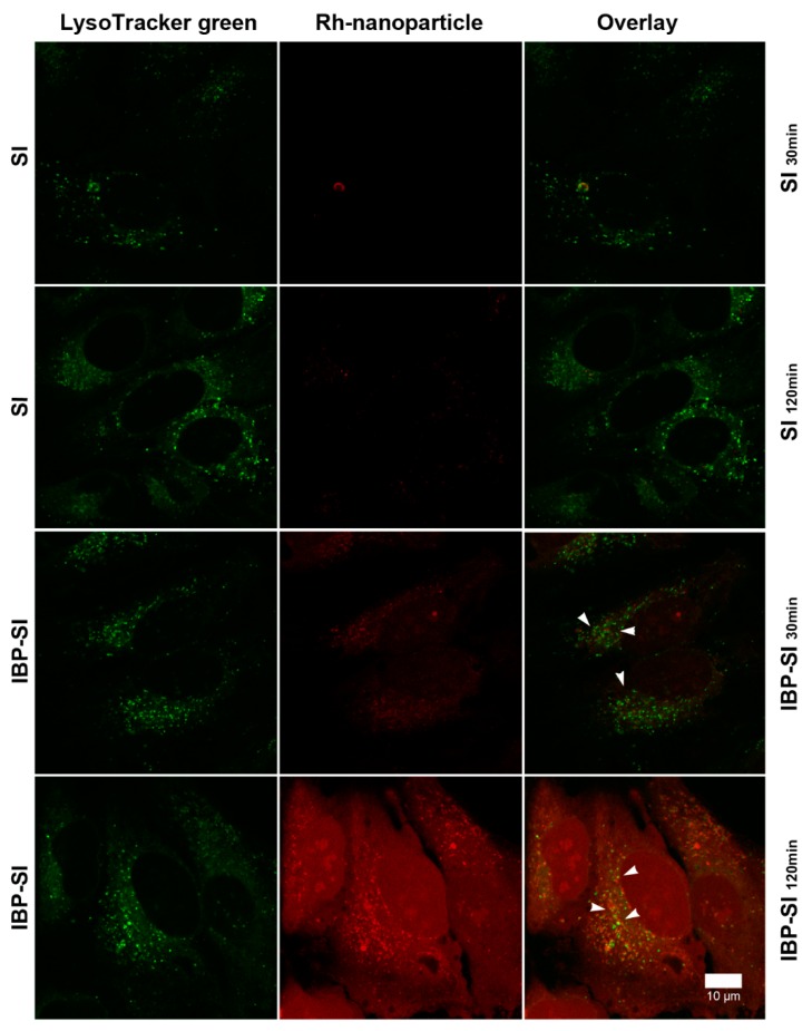
Figure A2.
IBP-SI exhibits a cross-species targeting activity to human ICAM-1. HeLa cells were treated with rhodamine-labelled ELPs at 37 °C for 30 min or 120 min prior to imaging using confocal fluorescence microscopy. LysoTracker Green (70 nM) was used to delineate low pH compartments. At comparable time points, IBP-SI exhibited more detectable internalization than SI. Green, low pH compartments (late endosomes and lysosomes); red, ELP nanoparticles; white arrowheads, internalized ELP nanoparticles. Scale bar = 10 µm.

