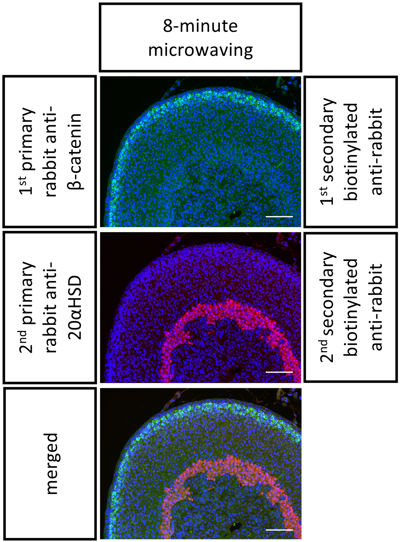
Figure 2. Representative double immunostaining on P21 FFPE mouse adrenals.

The FFPE mouse adrenal sections were stained with two primary antibodies from rabbits in the following order: anti-β-catenin (in green, stains the outer cortex) and then anti-20αHSD (in red, stains the inner cortex). An 8-min stripping step was performed in between. Scale bar 100 μm. DAPI stains cell nuclei, in blue.
