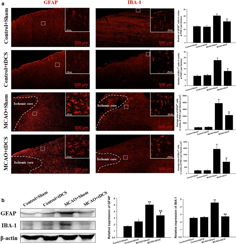
Fig. 5.
tDCS inhibited the activation of astrocyte and microglia induced by MCAO operation in CIP. a Immunohistochemistry (IHC) of GFAP+ and IBA-1+ cells; b The level and the semiquantitative analysis of GFAP and IBA-1. Data are presented as the mean ± SEM from at least three independent experiments, and representative images are shown. **P < 0.01, MCAO + Sham vs Control + Sham; ##P < 0.01 and #P < 0.05, MCAO + tDCS vs Control + tDCS; &&P < 0.01, MCAO + Sham vs MCAO + tDCS. Scale bar = 500 μm for macrographs and scale bar = 200 μm for micrographs

