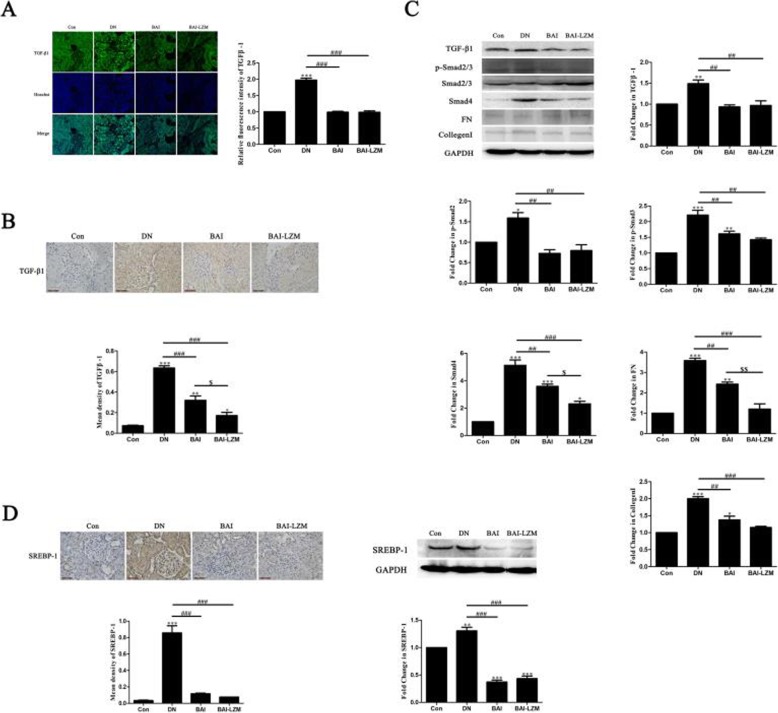
Fig. 6.
BAI-LZM inhibits extracellular matrix accumulation via the TGF-β1/Smad3 pathway in rats with DN. The protein level of TGF-β1 was detected by (A) immunofluorescence and (B) immunohistochemistry (magnification, × 400). (C) The protein levels of TGF-β1, phosphorylated-Smad2/3, Smad2/3, Smad4, FN and collagen I were detected by western blotting. The level of SREBP-1 was detected by (C) immunohistochemistry (magnification, × 400) and (D) western blotting. Con, saline; DN, 65 mg/kg STZ; DN/B, 65 mg/kg STZ + 160 mg/kg/day BAI; DN/B + L, 65 mg/kg STZ + 160 mg/kg/day BAI-LZM. The results are representative of 3 independent experiments. Data are presented as the mean ± standard deviation *P < 0.05, **P < 0.01 between the values in DN, DN/B and DN/B + L rats vs. the baseline levels (Con), as calculated by Student’s t test. #P < 0.05, ##P < 0.01 between the DN and BAI/BAI-LZM treatment groups, as calculated by one-way analysis of variance. $P < 0.05, $$P < 0.01 between the BAI and BAI-LZM treatment groups, as calculated by one-way analysis of variance. P-values were calibrated using the Bonferroni correction. BAI-LZM, baicalin-lysozyme; DN, diabetic nephropathy; STZ, streptozotocin; Con, control; TGF, transforming growth factor

