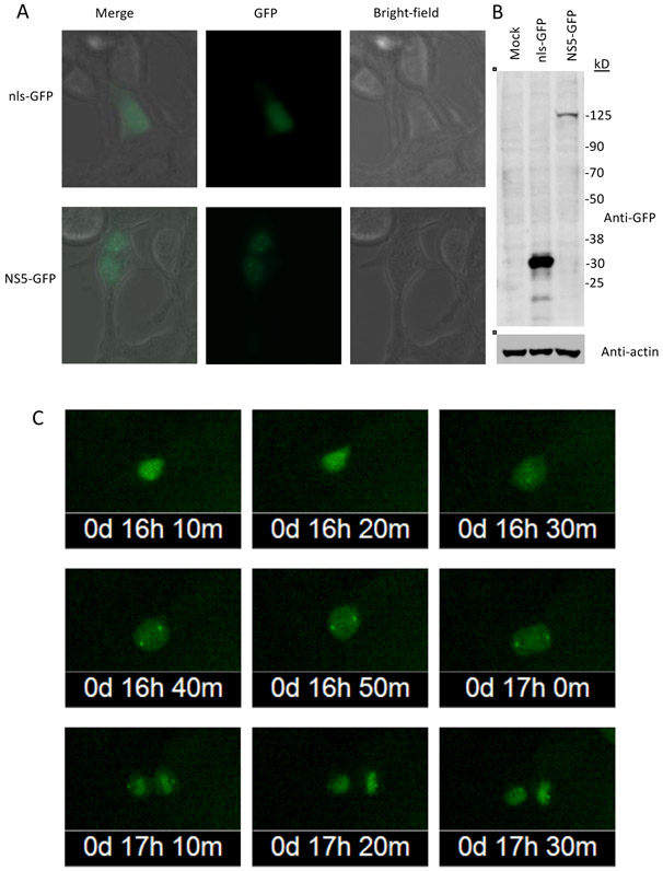
Fig. 3. Association of NS5-GFP with the centrosomes is maintained through mitosis.
HEK-293 cells were transiently transfected with NS5-GFP and nls-GFP expression plasmids.
A. NS5-GFP and nls-GFP expression were monitored by fluorescent microscopy (40X magnification).
B. Immunoblot analysis of NS5-GFP and nls-GFP expression. Blots were probed with anti-GFP antibodies. Molecular weight markers are shown at right (sizes in kDa). Predicted sizes of NS5-GFP and nls-GFP are 131 and 28 kDa, respectively.
C. Time-lapse images of an HEK-293 cell transiently expressing NS5-GFP undergoing mitosis.

