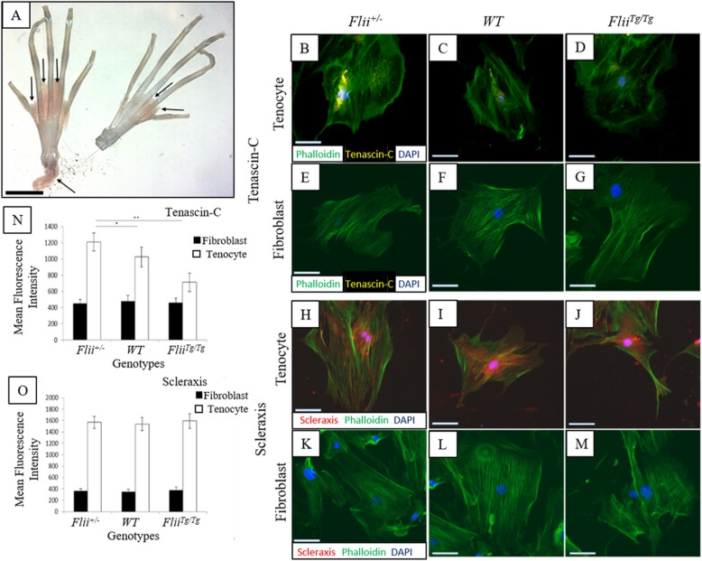
Fig. 1.
Tenascin-C and Scleraxis are expressed specifically in tenocytes. a Intact digital tendons were removed from the right and left hind paws of mice for tenocyte isolation. The tendon sheath is indicated by arrows and was removed before digestion to ensure a pure population of intrinsic tenocytes. Scale bar = 1 cm. Representative images of Tenascin C (b–g) and Scleraxis (h–m) staining in tenocyte and fibroblast cells isolated from Flii+/−, WT and FliiTg/Tg mice. Positive staining was detected in tenocyte cells for both Tenascin-C and Scleraxis, and no expression was noted in fibroblasts. Magnification × 20, scale bar = 100 μM. Graphical representation of mean fluorescence intensity of Tenascin C (n) and Scleraxis (o) staining. Tenascin-C staining was significantly higher in Flii+/− cells compared to WT and FliiTg/Tg cells. Data is represented as mean ± SEM. *p ≤ 0.05, **p ≤ 0.01, n = 6

