Fig. 4.
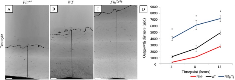
Differential effect of Flii expression on tenocyte outgrowth from tendon explants. 2 mm3 sections of tendons from Flii+/−, WT, and FliiTg/Tgmice were cultured for 12 days and tenocyte outgrowth measured as average migration distance. a–c Representative images of cellular outgrowth in Flii+/−, WT, and FliiTg/Tg tenocytes at 12 days post seeding. Dotted line + arrow = outgrowth distance. Magnification × 4. Scale bar = 200 μM. dFliiTg/Tg tenocytes have significantly increased outgrowth compared to WT and Flii+/− tenocytes. Data represented as mean ± SEM. *p ≤ 0.05, n = 6

