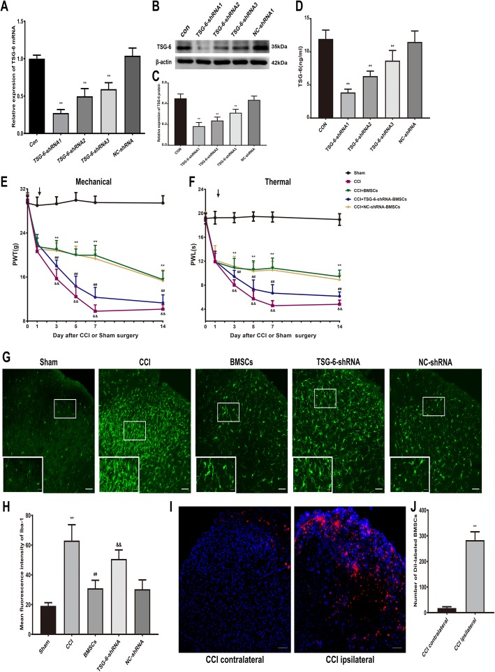
Fig. 5.
BMSCs release TSG-6 to inhibit neuropathic pain and suppress microglia activation in CCI rats. a–c The gene and protein expression of TSG-6 in control BMSCs, and in BMSCs transfected with TSG-6-shRNA1, TSG-6-shRNA2, TSG-6-shRNA3, and negative control (NC)-shRNA. d The ELISA analysis showed TSG-6 release in control BMSCs, and in BMSCs transfected with TSG-6-shRNA1, TSG-6-shRNA2, TSG-6-shRNA3, and negative control (NC)-shRNA. Reversal of BMSCs-induced inhibition of mechanical allodynia (e) and thermal hyperalgesia (f) in CCI rats by transfecting TSG-6-shRNA1. Arrows in (e, f) indicated the time of BMSCs injection. g Immunohistological staining showed that the inhibitory effect of BMSCs on microglia activation in the ipsilateral spinal cord dorsal horn was reduced after TSG-6 was knocked down. Scale bar: 50 μm; 10 μm (inserts). h Quantitative analysis of mean fluorescence intensity of Iba-1. i Localization of intrathecally injected CM-Dil-labeled BMSCs on day 3 after injection in the spinal cord dorsal horn. Scale bar: 50 μm. j Number of CM-Dil-labeled BMSCs in spinal cord dorsal horn. The data are expressed as the means ± SD (n = 8 in each group). **P < 0.01 versus the sham group. ##P < 0.01 versus the CCI + BMSCs group. &&P < 0.01 versus the BMSCs group. Statistical significance was determined by one-way analysis of variance (ANOVA) with post hoc Tukey test (a, c, d, h), two-way analysis of variance (ANOVA) with post hoc Tukey test (e and f), Student’s t test (two-tailed) (j)

