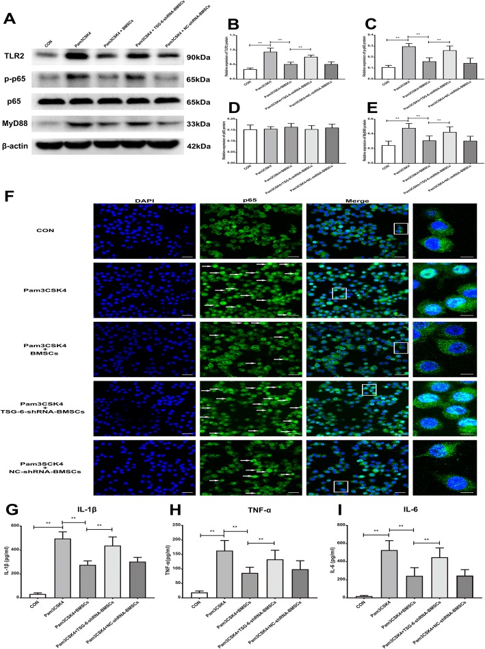
Fig. 9.
BMSCs released TSG-6 to reduce pro-inflammatory cytokine production through TLR2/MyD88/NF-κB signaling pathway in primary microglia. a Representative image of protein levels of TLR2, MyD88, p-p65, p65 in primary microglia. b–e Quantitative analysis of western blotting result showed that BMSCs co-culture inhibited the Pam3CSK4-induced the protein expression of TLR2, MyD88, p-p65 in primary microglia, in contrast, the inhibitory effect of BMSCs on the expression of these proteins was weakened after TSG-6 was knock down. f Typical micrographs of immunocytochemical staining are shown for the cytoplasmic and nuclear distribution of NF-κB p65. Scale bar: 30 μm; 10 μm (magnified graphs). g–i ELISA analysis showing BMSCs co-culture inhibited IL-1β, IL-6, and TNF-α release in Pam3CSK4-treated primary microglia; however, the anti-inflammatory effect of BMSCs was compromised after TSG-6-shRNA treatment. The data are expressed as the means ± SD (n = 8 in each group). **P < 0.01. Statistical significance was determined by one-way analysis of variance (ANOVA) with post hoc Tukey test

