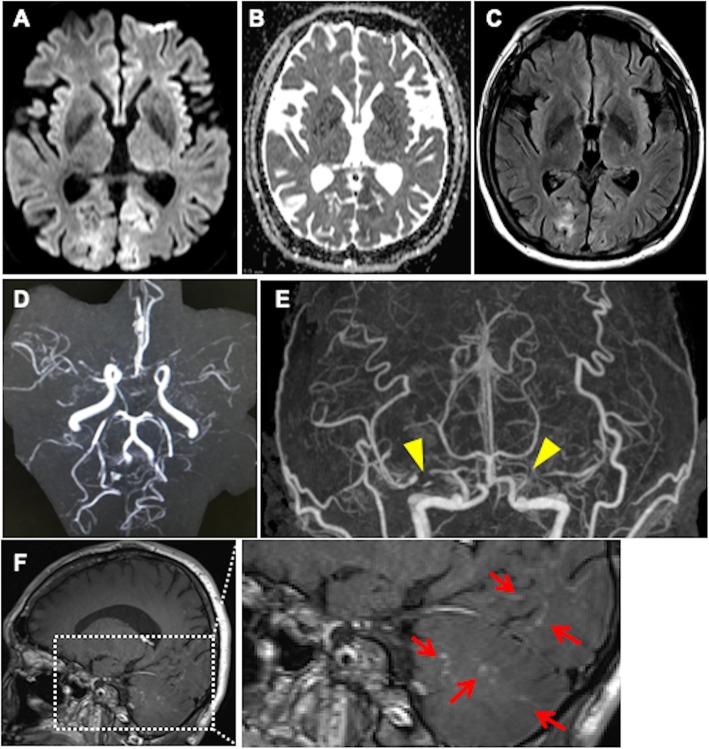
Fig. 2.
Radiological observations on the second admission a–c. Brain magnetic resonance imaging (MRI) on the second admission shows hyperintense lesions in the bilateral occipital lobe on diffusion-weighted imaging (a), apparent diffusion coefficient map (b), and fluid-attenuated inversion recovery (c). d. MR angiography shows stenosis in bilateral middle cerebral arteries (MCAs). e. Three-dimensional contrast-enhanced angiography revealed occlusions in bilateral MCAs (yellow arrowheads). f. Contrast-enhanced MRI shows multiple punctate and linear gadolinium-enhanced lesions (red arrows) in the occipital and temporal lobes and cerebellum

