Fig. 3.
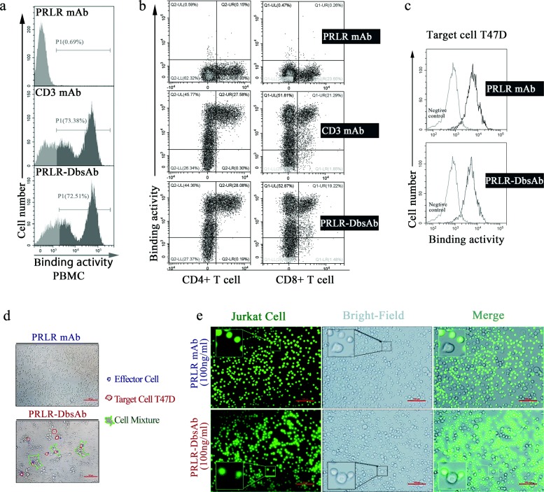
Purified PRLR-DbsAb redirects T cell to PRLR-expression T47D cells. PBMCs were incubated with 5 μg/ml PRLR-mAb, CD3 mAb or PRLR-DbsAb for 30 min and then the APC-labeled anti-human Fc secondary antibody were simultaneously added with FITC-labeled anti-CD8 and PE-labeled anti-CD4 antibodies (a and b). (a) FACS analysis of PRLR-DbsAb binding to isolated PBMCs. The grey region represents the negative fluorescent signal; the black region is the positive fluorescent signal. (b) Flow cytometry analysis of PRLR-DbsAb binding to CD4 and CD8 positive T lymphocytes. (c) Flow analysis histogram of PRLR-DbsAb binding PRLR-positive cell line T47D. The PE-labeled anti-human-Fc antibody was added after incubating with PRLR mAb and PRLR-DBsAb, followed by FACS fluorescence detection. It was observed respectively with a light microscope and a fluorescence microscope that the PRLR-DbsAb redirected T cells (d) and T cell-derived CD3-positive tumor Jurkat cells (e) to T47D cells. The experiment was repeated three times

