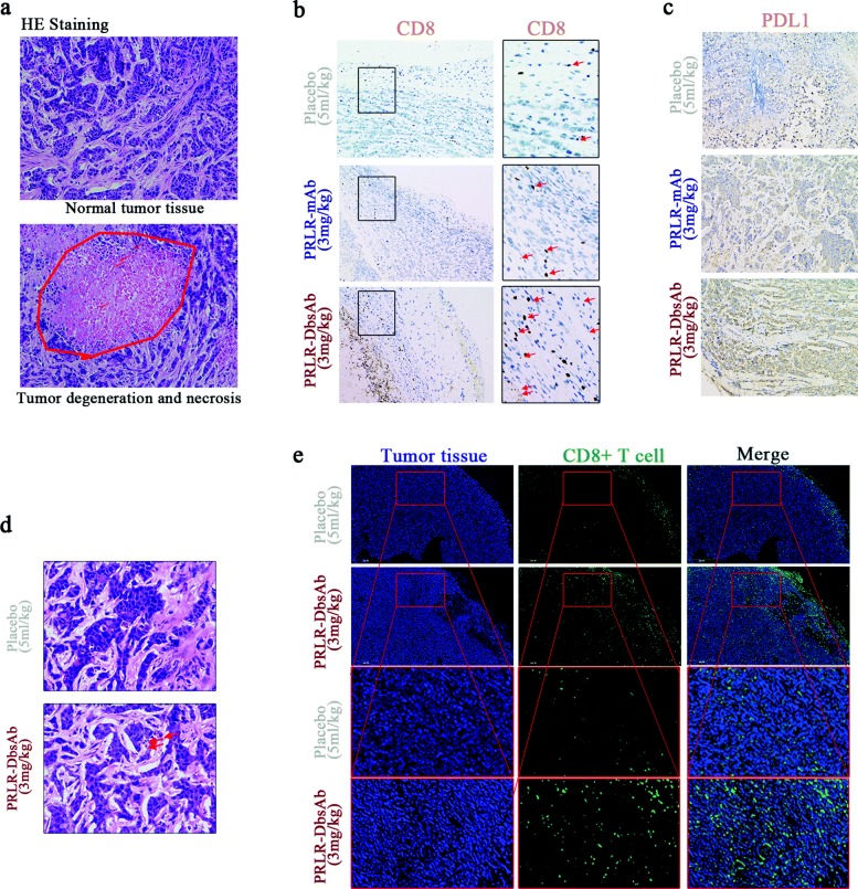
Fig. 6.
PRLR-DbsAb stimulates T cell infiltration and the PD-L1 expression unregulated in tumor tissue. sc Tumor cells plus sc effector cells (E/T 1:4) model: (a) HE staining of tumor tissue strapped from mice treated with 3 mg/kg PRLR-DbsAb. The above image showed normal tumor tissue, and the following image showed degenerate necrotic tumor tissue; (b) Immunohistochemical staining of CD8 in tumor tissue from the mice of different group; (c) Immunohistochemical staining of PD-L1 in tumor tissue from the mice of different group. Sc tumor cells plus ip effector cells (1:1) model: (d) HE staining; (e) Immunofluorescent staining of CD8. Three independent experiments were conducted and representative data is shown in this figure

