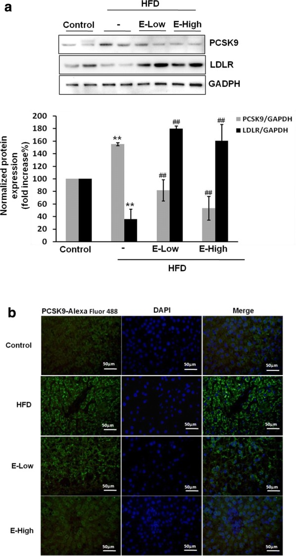
Fig. 2.

EGCG suppresses PCSK9 expression in hepatic tissue of rats. SD rats fed with HFD for 4 weeks and administered with different doses of EGCG by i.g. for another 4 weeks. a The hepatic protein samples were extracted and detected the expression of PCSK9 and LDLR by western blotting and densitomedtric analysis. b Hepatic tissue of rats were fixed in 4% paraformaldehyde and paraffin-embedded μm sections subjected to immunofluorescence analysis. Representative images of liver paraffin sections stained for PCSK9 with Alexa Fluor 488 (green) and counter stained with DAPI to show cell nucleus (blue). The photomicrographs were taken by Zeiss AX10 fluorescence microcopy at original magnification × 200. Results are representative of three independent experiments with similar results. **p < 0.01, vs control, ##p < 0.01, versus HFD group (N = 6). E-low: the lower dose of EGCG; E-high: the higher dose of EGCG
