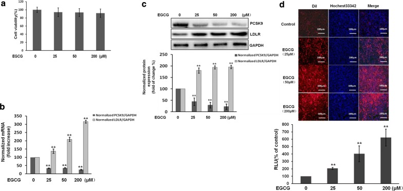
Fig. 3.
EGCG inhibits the expression of PCSK9, but promotes the LDLR expression and activity in HepG2 cells. The cells were treated with different doses of EGCG (25-200 μM) for 24 h. a Cell viability was measured using an MTS assay. b The mRNA expression of PCSK9 and LDLR were analyzed by real-time PCR. Relative expression changes are presented as the fold-increase of PCSK9/GADPH and LDLR/GAPDH. c The protein expression of PCSK9 and LDLR was analyzed by western blots. d Representative fluorescence microscopy images of cell‐associated Dil‐LDL (red), Hoechst‐stained nuclei (blue) and the overlay (upper). Fluorescence of isopropanol‐extracted Dil (520–570 nm, normalized to the cell protein) (lower). Data are presented as mean ± SEM (N = 3). *p < 0.05, **p < 0.01 compared with control

